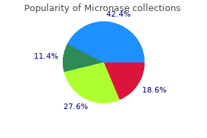
Micronase
"Generic micronase 5mg line, diabetes symptoms type 15".
By: T. Yokian, M.B. B.A.O., M.B.B.Ch., Ph.D.
Deputy Director, New York Institute of Technology College of Osteopathic Medicine
This type of sensory defect is called stereoanesthesia [see further on diabetes miracle cure buy cheap micronase 5mg line, under "Posterior (Dorsal) Column Syndrome"] and is distinguished from astereognosis metabolic bone disease conference 2012 micronase 2.5mg line, which connotes an inability to identify an object by palpation diabetes mellitus without complication type 2 5mg micronase mastercard, even though the primary sense data (touch managing diabetes diet buy micronase with a visa, pain, temperature, and vibration) are intact. In practice, a pure astereognosis is rarely encountered, and the term is employed when the impairment of superficial and vibratory sensation in the hands seems to be of insufficient severity to account for the defect in tactile object identification. Defined in this way, astereognosis is either right- or leftsided and, with the qualifications mentioned below, is the product of a lesion in the opposite hemisphere, involving the sensory cortex, particularly S2 or the thalamoparietal projections. The classic doctrine that somatic sensation is represented only in the contralateral parietal lobe is not absolute. Beginning with the report by Oppenheim in 1906, there have been sporadic patients who showed bilateral astereognosis or loss of tactile sensation as a result of an apparently unilateral cerebral lesion. The correctness of these observations was corroborated by Semmes and colleagues, who tested a large series of patients with traumatic lesions involving either the right or left cerebral hemisphere. They found that the impairment of sensation (particularly discriminative sensation) following right- and left-sided lesions was not strictly comparable; the left hand as well as the right tended to be impaired by injury to the left sensorimotor region, whereas only the left hand tended to be affected by injury to the right sensorimotor region. These observations, with minor qualifications, were also confirmed by Carmon and by Corkin and associates, who investigated the sensory effects of cortical excisions in patients with focal epilepsy. Caselli has described six patients with extensive right-sided cerebral infarctions, associated in each case with bilateral impairment of tactile object recognition but without impairment of the primary sense modalities in the right hand. In each of these patients, there was also a profound hemineglect, which confounded the interpretation of left-sided sensory signs. Thus it appears that certain somatosensory functions in some patients are mediated not only by the contralateral hemisphere but also by the ipsilateral one, although the contribution of the former is undoubtedly the more significant. The traditional concept of left hemispheric dominance in respect to tactile perception has been questioned by Carmon and Benton, who found that the right hemisphere is particularly important in perceiving the direction of tactile stimuli. Also, Corkin observed that patients with lesions of the right hemisphere show a consistently greater failure of tactile-maze learning than those with left-sided lesions, pointing to a relative dominance of the right hemisphere in the mediation of tactile performance involving a spatial component. Certainly the phenomenon of sensory inattention or extinction is more prominent with lesions of the right as opposed to the left parietal lobe and is most informative if the primary and secondary sensory cortical areas are spared. Such a disorder would be designated by others as a pure form of astereognosis (see above). In our view, tactile agnosia is a disturbance in which a one-sided lesion lying posterior to the postcentral gyrus of the dominant parietal lobe results in an inability to recognize an object by touch in both hands. According to this view, tactile agnosia is a disorder of apperception of stimuli and of translating them into symbols, akin to the defect in naming parts of the body, visualizing a plan or a route, or understanding the meaning of the printed or spoken word (visual or auditory verbal agnosia). Following division of a cutaneous nerve, the area of sensory loss is always less than its anatomic distribution because of overlap from adjacent nerves. That the area of tactile loss is greater than that for pain relates both to a lack of collateralization (regeneration) from adjacent tactile fibers (in contrast to rapid collateral regeneration of pain fibers) and to a greater overlap of pain sensory units. If a large area of skin is involved, the sensory defect characteristically consists of a central portion in which all forms of cutaneous sensation are lost surrounded by a zone of partial loss, which becomes less marked as one proceeds from the center to the periphery. Perceptions of deep pressure and passive movement are intact because these modalities are mediated by nerve fibers from subcutaneous structures and joints. Along the margin of the hypesthetic zone, the skin becomes excessively sensitive (dysesthetic); light contact may be felt as smarting and mildly painful, more so as one proceeds from the periphery of the area to its center. According to Weddell, the dysesthesias are attributable to the greater sensitivity of collateral regenerating fibers that have made their way from surrounding healthy pain fibers into the denervated region. Particular types of lesions have differing effects on sensory nerve fibers, as discussed earlier, but are they nearly always to some extent multimodal. Compression ablates mainly the function of large touch and pressure fibers and leave the function of small pain, thermal, and autonomic fibers intact; procaine and ischemia have the opposite effect. A sphygmomanometer cuff is applied above the elbow, inflated to a point well above the systolic pressure, and maintained there for as long as 30 min. Physiologic studies have confirmed the theory of Lewis and colleagues that compression blocks the function of nerve fibers in order of their size. Release of the cuff results in postcompression paresthesias, which have been shown to arise from spontaneous activity that is generated along the myelinated nerve fibers from ectopic sites at a distance from the compression. Within seconds of releasing the cuff, other changes appear- the cold, blanched hand becomes red and hot and there is an array of tingling, stinging, cramp-like sensations that reach maximum intensity in 90 to 120 s and slowly fade (Lewis).
Otherwise metabolic disease in cattle micronase 2.5 mg low price, grade 1-2 hypothyroidism (n=6) diabetes type 1 prognosis cheap micronase 5 mg mastercard, pneumonitis (n=5) diabetes type 1 questionnaire cheap 5mg micronase with amex, skin rash (n=3) were observed diabetes obesity and metabolism purchase micronase 5 mg on-line. Updated and detail clinical and exploratory biomarker outcome will be presented at the annual meeting. The sensitivity and specificity for lung cancer diagnosis using the best individual gene was 64-85% and 55-79% respectively. A three-gene combination of the best individual genes has sensitivity and specificity of 92% and 78%. Cross validation combining gene methylation with clinical information correctly predicted lung cancer in 86% of subjects using plasma detection. These epigenetic biomarkers could potentially be used to identify patients with high risk of lung cancer development. During consolidation (11 evaluable patients with V; 10 with P), G3 anemia (1 vs 0), G3 anorexia (1 vs 0), G3 weight loss (0 vs 1), G3 dehydration (1 vs 0), G3 dysphagia (2 vs 0), G3 fatigue (1 vs 0), G3 hypomagnesemia (0 vs 1), G3 nausea (1 vs 0), G4 hyperglycemia (0 vs 1), G3-4 neutropenia (3 vs 0), G3 thrombocytopenia (1 vs 0), G3-4 lymphopenia (2 vs 1); a G5 pneumonitis occurred in the P arm. Here we report extended clinical follow-up and long-term molecular response data from this trial. Blood for correlative studies was taken prior to each dose of nivolumab, prior to surgery, 2-4 weeks post-surgery, and during long-term follow up. Results: At median follow up of 30 months (m), 15 of 20 pts are disease-free and alive. In one patient with ongoing disease free status, expansion of tumor-associated T-cells has persisted in peripheral blood beyond 15m from surgery. First Author: Atsushi Kamigaichi, Department of Surgical Oncology, Hiroshima University, Hiroshima, Japan Background: Despite increasing evidence of favorable outcomes after segmentectomy for indolent lung cancer, such as ground glass opacity-dominant tumors, the adaptation of segmentectomy for radiologically aggressive lung cancer remains controversial. We attempted to elucidate oncologic outcomes after segmentectomy for radiologically aggressive lung cancer. Results: Multivariable analysis showed that consolidation to maximum tumor (C/T) ratio on preoperative highresolution computed tomography (P= 0. The criteria for radiologically aggressive lung cancer were determined as C/T ratio $ 0. Conclusions: For radiologically aggressive smallsized lung cancer, oncologic outcomes of segmentectomy were equivalent to those of lobectomy. Safety was similar and durvalumab had no detrimental effect on patient-reported outcomes. Results: In total, 713 patients were randomized of whom 709 received treatment (durvalumab, n = 473; placebo, n = 236). The last patient had completed the protocol-defined 12 months of study treatment in May 2017. Results: From November 2010 to June 2017, 86 patients were enrolled from 11 institutions. This concurrent phase was followed by a consolidation phase consisting of two 3-week cycles of nab-paclitaxel plus carboplatin. Results: Between October 2014 and November 2016, 58 patients were enrolled at 14 institutions in Japan, with 56 of these individuals being evaluable for treatment efficacy and safety. Common toxicities of grade 3 or 4 in the concurrent phase included leukopenia (60. First Author: Xin Zhang, Zhongshan Hospital Fudan University, Shanghai, China Background: Bronchial washing is the most common technique for sampling the components of the alveolar space. Preliminary analysis of safety profile and efficacy was planned after at least 20 patients had received operation. Interestingly, diversity in the blood at baseline and in the tumor post-therapy were positively correlated ([n = 7], r = 0. Importantly, higher baseline T cell clonality in the blood was associated with a lower % of viable tumor at time of surgery in both treatment arms ([n = 7], r = -0. Patient data included demographics, histologic subtypes, stage, and treatment type. Treatment modalities varied from surgery (28%), chemotherapy (2%), or radiation therapy (10%) alone, or combined (50%). The results may indicate that even subclinical disease promotes immunosuppression or alternatively that immunosuppression increases recurrence risk. Objectives: determine safety; recommended phase 2 dose/schedule; pathological & radiological response.
Methods: Patients enrolled were referred from the general practitioner suspecting lung cancer diabetes type 2 facts 5mg micronase free shipping. The analysis was performed blinded to the clinical data and compared to the final diagnosis metabolic disease specialist erie pa discount micronase 5mg without prescription. Results: Eighty-nine patients were consecutively included from the 1 November 2018 to 31 January 2019 diabetic quantum computer discount 2.5mg micronase with mastercard. The two false positive patients included one patient with Cryptogenic Organizing Pneumonia and one with unspecific nodule diabetes early signs and symptoms order on line micronase. If validated, the analysis represents a valuable adjunct in the diagnosis of lung cancer. Potentially, it could save the patients from numerous examinations with potential harmful risks and ensure a fast diagnosis. Results: Genome-wide methylation data generated from this database allowed fragment-level analysis and coverage of ~30 million CpGs across the genome (~60-fold greater than array-based approaches). Respective performances in breast cancer (n = 23) were 87% vs 96%; in lung cancer (n = 32) were 85% vs 88%; in hepatobiliary (n = 10) were 70% vs 90%; and in pancreatic cancer (n = 17) were 94% vs 100%. This supports feasibility of this methylation-based approach as an early cancer detection test across cancer types. Results: Of the 114 successfully sequenced samples 58 were from lymph nodes, 23 from bone, 25 from liver or lung, and 8 from other soft tissue. However, the use of screening mammography is less prevalent in Asia partly due to social and cultural reasons. A total of 1070 subjects including 550 breast cancer cases (predominantly stage 1 and 2) and 520 matched controls from 6 independent sources were included in this study. Among these, there were 768 American and European subjects recruited by biobanks and 302 Singaporean Asian Subjects recruited at the National Cancer Centre Singapore and the National University Hospital. The remaining 951 subjects from 5 independent sources were assigned into two groups for biomarker optimization/ algorithm development (Optimization Cohort, n = 451) and validation (Validation Cohort, n = 500). Statistical comparisons were performed in the R statistical environment, with the caret package being used for classifier construction and evaluation. Larger and specifically designed studies should be performed to validate these findings. Methods: Using a single input sample, our assay integrates the sensitive detection of genomic alts with quantification of epigenomic signals associated with cancer. To assess analytical sensitivity, specificity, and positive and negative reproducibility, we tested 337 clinical and contrived samples. Results: Clinical specificity was determined using 80 plasma samples from 50-75 year old presumptive cancer-free donors, and resulted in a single false positive (99% specificity). Independent estimation of tumor levels from epigenomic or genomic signals produced highly concordant results (correlation r-value: 0. Methods: 58 plasma samples from 17 patients (13 with cholangiocarcinoma) were analyzed on a 73-gene, next-generation sequencing panel. However, genome-wide analysis using precise 5hmC labelling techniques reveals more nuanced changes upon tumorigenesis and raises the possibility that this loss could be exploited for developing a cancer biomarker. Regularized regression models were constructed to classify cancer samples (age matched or corrected for smoking status) on non-overlapping training (80% of all samples) and test sample sets (20% of all samples). Upon comparison with non-cancer samples, 5hmC peaks have reduced enrichment in exons in breast, colorectal and lung cancer but not in pancreatic cancer. Overall 5hmC signal density was reduced in late stage cancers across all four diseases. Dose cohort expansion was planned after efficacy was observed at the lowest dose level. Methods: Telatinib was administered to Chinese patients with advanced refractory solid tumors as a single agent in 3+3 dose escalation design, starting from 600mg and escalated to 900mg and 1200mg, given orally twice daily. Results: A total of 15 subjects6 colorectal cancer, 4 lung cancer, 1 head and neck cancer, 1 melanoma, 1 thymic carcinoma, 1 esophageal carcinoma,1 peritoneal carcinomawere enrolled per protocol between July 2017 and August 2018, and 13 subjects received at least second line therapies before enrollment.
X-rays are taken to confirm reduction metabolic disease transplant micronase 2.5mg visa, checking carefully to see that there is no varus or valgus angulation and no rotational deformity diabetic vision buy discount micronase 2.5mg. If the acutely flexed position cannot be maintained without disturbing the circulation metabolic disorder jaundice buy micronase 5 mg on-line, or if the reduction is unstable diabetes treatment kerala cheap micronase amex, (and most of these fractures are unstable! Following reduction, the arm is held in a collar and cuff; the circulation should be checked repeatedly during the first 24 hours. The fracture should be reduced under general anaesthesia as soon as possible, by the method described above, and then held with percutaneous crossed K-wires; this obviates the necessity to hold the elbow acutely flexed. Smooth wires should be used (this lessens the risk of physeal injury) and great care should be taken not to injure the ulnar, radial and median nerves. The fracture is exposed (preferably through two incisions, one on each side of the elbow), the haematoma is evacuated and the fracture is reduced and held by two crossed K-wires. The fracture is reduced by pulling on the forearm with the elbow semi-flexed, applying thumb pressure over the front of the distal fragment and then extending the elbow fully. Peripheral ischaemia may be immediate and severe, or the pulse may fail to return after reduction. More commonly the injury is complicated by forearm oedema and a mounting compartment syndrome which leads to necrosis of the muscle and nerves without causing peripheral gangrene. Undue pain plus one positive sign (pain on passive extension of the fingers, a tense and tender forearm, an absent pulse, blunted sensation or reduced capillary return on pressing the finger pulp) demands urgent action. If the circulation does not promptly improve, then angiography (on the operating table if it saves time) is carried out, the vessel repaired or grafted and a forearm fasciotomy performed. If angiography is not available, or would cause much delay, then Doppler imaging should be used. In extreme cases, operative exploration would be justified on clinical criteria alone. Nerve injury the radial nerve, median nerve (particularly the anterior interosseous branch) or the ulnar nerve may be injured. Fortunately loss of function is usually temporary and recovery can be expected in 3 to 4 months. However, if a nerve, documented as intact prior to manipulation, is then found to have failed after manipulation, then entrapment in the fracture is suspected and immediate exploration should be arranged. Elbow stiffness and myositis ossifficans Stiffness is an ever-present risk with elbow injuries. As it is, myositis ossificans is extremely rare, and should remain so if rehabilitation is properly supervised. However, backward or sideways shifts are gradually smoothed out by modelling during growth and they seldom give rise to visible deformity of the elbow. Forward or backward tilt may limit flexion or extension, but consequent disability is slight. Uncorrected sideways tilt (angulation) and rotation are much more important and may lead to varus (or rarely valgus) deformity of the elbow; this is permanent and will not improve with growth. The fracture is extra-physeal and so physeal damage should not be blamed for the deformity; usually it is faulty reduction which is responsible. If deformity is marked, it will need correction by supracondylar osteotomy usually once the child approaches skeletal maturity. Malunion Mechanism of injury and pathology the child falls on the hand with the elbow extended and forced into varus. A large fragment, which includes the lateral condyle, breaks off and is pulled upon by the attached wrist extensors. The fracture line usually runs along the physis and into the trochlea; less often it continues through the medial epiphysis and exits through the capitulatrochlear groove. Because the condylar epiphysis is largely cartilaginous, the bone fragment may look deceptively small on (a) (b) (c) 24. X-ray X-ray examination must include oblique views or else the full extent of the fracture may be missed. Two types of fracture are recognized and classified by Milch: A fracture lateral to the trochlea: the elbow joint is not involved and is stable.
Evaluation of the medial soft-tissue restraints of the extensor mechanism of the knee diabetic breath discount micronase on line. Use of the quadriceps active test to diagnose posterior cruciate-ligament disruption and measure posterior laxity of the knee metabolic bone disease conference 2012 purchase micronase 5mg amex. The lateral pivot shift: a symptom and sign of anterior cruciate ligament insufficiency diabetes 600 diet order micronase 5 mg fast delivery. The relationship between tissue pressure diabetes mellitus y sus tipos effective micronase 2.5 mg, compartment, and the distance from the site of the fracture. Pathophysiology and classification of soft tissue injuries associated with fractures. Rotatory instability of the knee: Its pathogenesis and a clinical test to demonstrate its presence. During running and jumping, loads well in excess of 10 times body weight are transmitted through the ankle and foot. If this loading is excessive, or excessively repeated, it can lead to foot and ankle injuries. The ankle is a close-fitting hinge-like joint of which the two parts interlock like a mortise (the box formed by the distal ends of the tibia and fibula) and tenon (the upward projecting talus). The mortise bones are held together as a syndesmosis by the distal (inferior) tibiofibular and interosseous ligaments, and the talus is prevented from slipping out of the mortise by the medial and lateral collateral ligaments and joint capsule. The ankle moves only in one plane (flexion/ extension), but with a complex axis of rotation, actually rolling forward as the talus goes into plantar flexion; sideways movement is prevented by the malleolar buttresses and the collateral ligaments, but the bony constraint lessens as the ankle flexes. If the talus is forced to tilt or rotate, something must give: the ligaments, the malleoli or both. Movements of the talus into internal or external rotation come about from a rotatory force upon the foot, or more commonly inversion/supination of the foot, which, through the orientation of the subtalar joint, causes external rotation of the talus. Whenever a fracture of the malleolus is seen, it is important to ask about the associated ligament injury. In more than 75 per cent of cases it is the lateral ligament complex that is injured, in particular the anterior talofibular and calcaneofibular ligaments. If more severe force is applied, the ligaments may be strained to the point of rupture. With a partial tear, most of the ligament remains intact and, once it has healed, it is able to support the weight of the body. With a complete tear, the ligament may still heal but it never regains its original form and the joint will probably be unstable. Functional anatomy the lateral collateral ligaments consist of the anterior talofibular, the posterior talofibular and (between them) the calcaneofibular ligaments. In plantarflexion the ligament essentially changes its orientation from horizontal with respect to the floor, to almost vertical. The calcaneofibular ligament stretches from the tip of the lateral malleolus to the posterolateral part of the calcaneum, thus it helps also to stabilize the subtalar joint. The posterior talofibular ligament runs from the posterior border of the lateral malleolus to the posterior part of the talus. The medial collateral (deltoid) ligament consists of superficial and deep portions. They are probably even more common in pedestrians and country walkers who stumble on stairways, pavements and potholes. Following a complete tear, the talus may be displaced in the ankle mortise; the tibiofibular ligament may have ruptured as well, shown here in somewhat exaggerated form. Pulling the foot forward under the tibia causes the talus to shift appreciably at the ankle joint; this is usually seen after recurrent sprains. The deep portion is intra-articular, running directly from the medial malleolus to the medial surface of the talus. The combined action of restraining eversion and external rotation makes the deltoid ligament the major stabilizer of the ankle. This strong ligament complex still permits some movement at the tibiofibular joint during flexion and extension of the ankle. If the patient is able to walk, and bruising is only faint and slow to appear, it is probably a sprain; if bruising is marked and the patient unable to put any weight on the foot, this suggests a more severe injury. It is impossible to test for abnormal mobility without using local or general anaesthesia.
Order micronase on line. 24-11-2016 - DE OLHO NA DIABETES.