
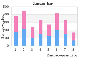
Zantac
"Generic zantac 150 mg with amex, dukan diet gastritis".
By: E. Tjalf, M.B. B.CH. B.A.O., M.B.B.Ch., Ph.D.
Deputy Director, Lake Erie College of Osteopathic Medicine
In the initial phases of cirrhosis chronic gastritis joint pain buy generic zantac 300 mg on line, compensation occurs through the development of hyperdynamic circulation (high plasma volume gastritis pronounce trusted 300mg zantac, cardiac index gastritis acid reflux diet order zantac 150mg line, and heart rate); however gastritis causes cheap zantac 300mg amex, as cirrhosis progresses and splanchnic arterial vasodilation increases, this compensatory mechanism is insufficient to maintain circulatory homeostasis. The forward theory of ascites formation follows from the peripheral arterial vasodilation hypothesis and holds that arterial vasodilation in the splanchnic circulation induces the formation of ascites by simultaneously impairing both the systemic circulation (leading to sodium and water retention) and the splanchnic microcirculation (where the forward increase in capillary pressure and permeability from the greatly increased inflow of blood at high pressure into the splanchnic capillaries leads to the leakage of fluid into the abdominal cavity). More recently, it has been suggested that the pathogenesis of circulatory dysfunction in cirrhosis is even more complex than that just described, such that the primary mechanism behind impaired circulatory function is worsening splanchnic arterial vasodilation that occurs in parallel with the progression of liver disease. It appears that cirrhosis is accompanied by a progressive impairment in both cardiac inotropic and chronotropic functions. The net effect of these abnormalities is a reduction in cardiac output and a decrease or disappearance of the hyperdynamic circulation. The mechanism of cardiac dysfunction in cirrhosis is not well established, but it is probably multifactorial. All these mechanisms may account for the progressive decrease in cardiac output observed in advanced cirrhosis. Therefore, the clinical course of cirrhosis can be divided into phases according to the onset of each of these complications (Box 30. Although patients remain able to excrete dietary sodium, subtle abnormalities in renal sodium excretion are present. For example, these patients have a reduced natriuretic response to the acute administration of sodium chloride. Abnormal natriuretic responses to changes in posture can also be seen during this phase: urinary sodium excretion is reduced in the upright and increased in the supine posture, compared with normal subjects. Their course is usually progressive except that, in patients with alcoholic cirrhosis, kidney function may improve after alcohol withdrawal. The main consequence of impaired sodium excretion in cirrhosis is the development of sodium retention and ascites. This occurs when renal sodium excretion decreases to less than the sodium intake and represents a marked impairment in renal sodium handling. Urinary sodium excretion, although reduced, is usually greater than 10 mEq/day and in some cases it exceeds 50 to 90 mEq/day; therefore, dietary sodium restriction may be sufficient to effect negative sodium balance and loss of ascites. Furthermore, plasma levels of atrial natriuretic peptide, brain natriuretic peptide, and natriuretic hormone are increased in these patients, indicating that sodium retention is not caused by a reduced synthesis of endogenous natriuretic peptides. No studies have compared the systemic hemodynamics in patients with portal hypertension and compensated cirrhosis with those in patients at this phase of the disease. However, two studies in the latter group of patients clearly showed the presence of hyperdynamic circulation with high cardiac output, low peripheral vascular resistance, and hypervolemia. Therefore, severely impaired kidney function may occur at this phase if renal prostaglandins are inhibited with the use of nonsteroidal antiinflammatory drugs. The International Ascites Club considers that serum creatinine concentration should be greater than 1. The arterial vascular resistance in these patients is increased not only in the kidneys, but also in the brain, muscle, and skin, indicating generalized arterial vasoconstriction in an attempt to compensate for intense splanchnic arterial vasodilation. The delivery of sodium to the distal nephron (where most diuretics act) is very low; therefore, most of these patients do not respond to diuretics and have refractory ascites. Free water clearance is also markedly reduced, and most patients have significant hyponatremia. Therefore, sodium retention in patients at this phase is caused by increased sodium reabsorption throughout the nephron. The plasma volume and peripheral vascular resistance do not differ from those observed in the previous phase. In fact, kidney, cerebral, and muscle blood flow in cirrhosis correlates inversely with plasma renin activity and the concentration of norepinephrine. The most interesting feature is that cardiac output in patients at phase 3 (although higher than in normal subjects) is lower than in patients at phase 2, indicating that progression of circulatory dysfunction is caused not only by an increase in splanchnic arterial vasodilation but also by a decrease in cardiac output. Patients succumb from progressive circulatory, hepatic, and kidney failure, along with encephalopathy. It is unknown whether this decrease in heart function is caused by cirrhotic or sepsis-related cardiomyopathy, by decreased cardiac preload resulting from central hypovolemia, or by both.
With this arrangement gastritis symptoms chest pain buy zantac 150mg with amex, it forms an active and dominant role in both storage and voiding gastritis snacks purchase generic zantac online. Inner longitudinal - It courses downwards from the fundus of the bladder and continues in the form of spirals upto the midurethra gastritis bloating buy 150 mg zantac with amex. A distal one which varies little with age and a proximal one beneath the bladder neck which undergoes marked changes with age gastritis symptoms remedy purchase 150 mg zantac with visa. In the reproductive period, these vessels give a cavernous appearance to the submucosa which disappears in the postmenopausal period. This urethral vascular system plays a significant role in the maintenance of resting urethral pressure. Support to bladder neck and urethra Support is maintained by intrinsic and extrinsic factors. Extrinsic factors: (i) Contraction of pubococcygeus part of levator ani muscle; (ii) Pubourethral ligaments and condensed endopelvic fascia with smooth muscle fibres; (iii) Exercise to increase collagen turnover and also to maintain strength of levator ani. Recent studies, however, suggest that there is frequent interchange of fibers between the bundles and the separate layers are not distinctly defined. From a functional point of view, the detrusor appears to contract as a single syncytial mass. The detrusor muscles are shown to contain significant amount of acetylcholinesterase. This part has got an additional support by the intrinsic striated muscle (rhabdosphincter urethrae). This muscle encircles the whole urethra and is composed predominantly of skeletal muscle with nerve supply from parasympathetic division of autonomic nerves. This rhabdosphincter is further enforced in the upper part by levator ani muscles (extrinsic muscles) being separated from it by a distinct connective tissue septum. The extrinsic periurethral muscle (levator ani) is supplied by the perineal branch of pudendal nerve. The intrinsic striated muscles (slow twitch fibers) is responsible for urethral closure at rest. The extrinsic periurethral striated muscles (first twitch fibers) provide additional support to urethra on stress. Distal urethra: this part is a passive conduit and is surrounded by collagen tissue. Pubourethral ligaments and condensed endopelvic fascia are found to contain smooth muscle fibers. They work together to maintain the normal anatomic support and prevent hypermobility of bladder neck and urethra. Preganglionic sympathetic fibers arising from T10 - L2 are also cholinergic but the postganglionic fibers innervating both the bladder and urethra act through the release of norepinephrine (adrenergic nerve fibers). The former component relaxes both the bladder and urethra and latter one contracts only the urethra. The sympathetic is concerned mainly with the filling and storage phase of micturition. Parasympathetic supply (acetylcholine) is responsible for detrusor contraction and normal voiding. The rhabdosphincter is supplied by pelvic splanchnic nerves traveling with the parasympathetic fibers. Extrinsic periurethral striated muscle is supplied by the motor fibers of the pudendal nerves. The intravesical pressure is raised to remain at almost steady level of about 10 cm of water even with a volume of about 500 ml. The intravesical pressure is kept lower than that of the urethra by delicately coordinated relaxation of detrusor muscle. Proximal urethral musculature acts like a sphincter by maintaining tonic contraction. Stretching of the detrusor reflexly contracts the sphincteric muscles of the bladder neck. Inhibition of the cholinergic system responsible for detrusor contraction operating from the spinal centers. The other component of the external sphincter derived from the levator ani, composed of fibres of "first twitch" variety innervated by the perineal branch of pudendal nerve.
Buy zantac 150mg mastercard. Home Remedies For Nasal Polyps II नाक की बीमारी का घरेलु उपचार II.
However all these grafts require a second surgical intervention and are associated with donor site morbidity gastritis diet soy milk purchase discount zantac online. Our study compared the "kazanjian technique without collagen" and "kazanjian technique with collagen" gastritis pediatric symptoms order 150mg zantac otc. All the patients showed excellent compatibility to collagen gastritis zungenbrennen purchase zantac, there was one incidence of infection gastritis diet of worms buy zantac paypal, and in one case there was excess increase in the vestibular depth due to early breakdown of collagen. Patients were referred to Department of Prosthodontics for early denture construction around 4 weeks to maintain the newly developed vestibular depth. Pre prosthetic vestibular sulcus extension by operation of Edlan and Mejchar, Int J oral surg 1979; 8: 333 339. A patient survey of denture tolerance before and after a mandibular vestibuloplasty with skin grafting. Influence of skin graft pathology on residual ridge reduction after mandibular vestibuloplasty. Saffa, Use of amnion as a graft material in vestibuloplasty: a preliminary report. Hydroxyapatite augmentation of mandible with simultaneous mucosal graft vestibuloplasty. The present study was a correlation between 24 hour urine protein excretion and random urine protein: creatinine ratio in diabetic subjects for predicting renal involvement. It was observed that protein excretion in 24 hr sample correlated significantly with random urine protein: creatinine ratio in physiological and also in nephrotic range of proteinuria. This may have important clinical applications as single urine specimens, which can be collected easily in outpatient clinics and field studies, could replace the more traditional timed urine collections that have been used to assess the risk of clinical diabetic renal disease. Occurrence of nephropathy is around 40% in type 1 and 25% in type 2 diabetes mellitus1. Degree of proteinuria is the useful marker for renal involvement and response to treatment. Random urine sampling for protein:creatinine ratio would be more acceptable and less time consuming. The protein:creatinine ratio takes into account the fact that creatinine excretion remains fairly constant in presence of a stable Glomerular Filtration Rate. Therefore the ratio of the two in a single voided sample would reflect the cumulative protein excretion over the day, as the two stable rates would cancel out the time factor2. The protein:creatinine ratio in a single voided urine samples is an accurate, convenient, inexpensive and reliable estimate of total proteinuria in the vast majority of patients. Hence protein: creatinine ratio in single voided urine samples may be even more reproducible than 24 hours urinary protein excretion3. The protein: creatinine ratio of a randomly obtained urine specimen correlates with 24-hour urine protein in patients with type 1 diabetes and may be a useful tool in screening a patient for proteinuria or estimating range of proteinuria in patients 4. Detailed medical history and relevant clinical examinations were carried out in these patients. Patients with chronic renal failure, glomerular nephritis due to other systemic conditions and hypertensives were excluded from the study. This was thoroughly mixed and a sample of 2ml was taken for evaluation of proteins. A random urine sample of 5ml was collected on next day any time just before the analysis. It is evident that correspondence of 24 hr urine protein and protein:creatinine ratio in diabetic subjects is highly significant (p<0. Similarly those patients who excreted < 200 mg protein in 24 hour had protein:creatinine ratio of < 0. Hence a highly significant correlation exist between 24-hour urinary protein concentration and protein: creatinine ratio levels in random urine sample 10. By careful choice of cut-off, protein: creatinine ratio can be used in patients with kidney disease to rule out abnormal 24 hr loss of protein. The definitive method for quantifying urinary protein excretion is 24-hour urine sample, but obtaining these samples is cumbersome, time consuming and inaccurate. The present study suggests estimating protein: creatinine ratio in random urine sample for renal involvement in diabetic subjects provide a convenient method for early diagnosis and intervention of diabetic nephropathy. Quantitation of proteinuria by the use of protein to creatinine ratios in single urine samples. The urine protein to creatinine ratio as a predictor of 24-hour urine protein excretion in type 1 diabetic patients with nephropathy.
The right pupillary constriction associated with light directed at the left eye requires the left optic nerve gastritis diet forum best 150mg zantac. The spinal part of the accessory nerve supplies the trapezius muscle gastritis diet ���������� zantac 150 mg with mastercard, which shrugs the shoulder gastritis diet japan buy zantac american express. The trigeminal nerve supplies the muscles of mastication responsible for chewing (see p definition de gastritis zantac 150mg discount. The facial nerve receives the sensation of taste from the anterior two-thirds of the tongue. The glossopharyngeal nerve receives the sensation of touch from the posterior third of the tongue (see p. Internal ophthalmoplegia is a condition in which the oculomotor nerve supply to the sphincter pupillae and the ciliary muscle is lost, but the innervation of the extraocular muscles is spared (see p. External ophthalmoplegia is a condition in which the oculomotor nerve supply to the extraocular muscles is lost, but the innervation of the sphincter pupillae and the ciliary muscle is spared (see p. The optic nerve leaves the orbital cavity through the optic canal in the lesser wing of the sphenoid bone. The olfactory receptor cells are located in the mucous membrane of the nasal cavity above the level of the superior concha (see p. The main sensory nucleus of the trigeminal nerve lies in the brainstem lateral to the motor nucleus. Proprioceptive impulses from the facial muscles end in the mesencephalic nucleus of the trigeminal nerve (see p. The facial nerve leaves the posterior cranial fossa with the vestibulocochlear nerve and enters the internal acoustic meatus. The superior salivatory nucleus of the facial nerve innervates the submandibular and sublingual salivary glands (see p. The taste sensation from the mucous membrane covering the anterior two-thirds of the tongue is conducted in the facial nerves and the chorda tympani nerves, which are a considerable distance from the metastases in the deep cervical lymph nodes in the neck. The wasted right half of the tongue and the pointing of the protruded tongue to the right side indicated a lesion of the right hypoglossal nerve. The tongue muscles on the right side had atrophied and diminished in size,resulting in the wrinkling of the overlying normal mucous membrane. The answers to questions 15 through 23 pertain to Figure 11-26, which shows the inferior view of the brain. The answers to questions 24 through 29 pertain to Figure 11-27, showing a medial view of the right side of the brain following a median sagittal section. Structure number 1 is the nucleus of the oculomotor nerve in the tegmentum of the midbrain at the level of the superior colliculus. Structure number 2 is the nucleus of the trochlear nerve in the tegmentum of the midbrain at the level of the inferior colliculus. Structure number 3 is the trigeminal nerve emerging on the anterior surface of the pons. Structure number 4 is the oculomotor nerve emerging from the anterior surface of the midbrain in the interpeduncular fossa. Patterns of sensory loss following fractional posterior fossa Vth nerve section for trigeminal neuralgia. The neuropsychology of facial expression: A review of the neurological and psychological mechanisms for producing facial expressions. The patient was conscious and was unable to feel any sensation down the right side of his body. There was no evidence of paralysis on either side of the body,and the reflexes were normal. Three days later, the patient appeared to be improving, and there was evidence of return of sensation to the right side of his body. The patient, however, seemed to be excessively sensitive to testing for sensory loss. On light pinprick on the lateral side of the right leg, the patient suddenly shouted out because of excruciating burning pain, and he asked that the examination be stopped. Although the patient experienced very severe pain with the mildest stimulation, the threshold for pain sensitivity was raised, and the interval between applying the pinprick and the start of the pain was longer than normal; also, the pain persisted after the stimulus had been removed. Moreover, the patient volunteered the information that the pain appeared to be confined to the skin and did not involve deeper structures.
If the patient is excessively sensitive to sound in one ear gastritis diet ��� order zantac 150 mg with mastercard, the lesion probably involves the nerve to the stapedius muscle gastritis diet ������� zantac 300mg free shipping, which arises from the facial nerve in the facial canal gastritis diet shopping list order zantac 150mg with mastercard. Loss of taste over the anterior two-thirds of the tongue indicates that the facial nerve is damaged proximal to the point where it gives off the chorda tympani branch in the facial canal gastritis turmeric generic 300mg zantac with amex. A firm swelling of the parotid salivary gland associated with impaired function of the facial nerve is strongly indicative of a cancer of the parotid gland with involvement of the nerve within the gland. The part of the facial nucleus that controls the muscles of the upper part of the face receives corticonuclear fibers from both cerebral hemispheres. Therefore, it follows that with a lesion involving the upper motor neurons,only the muscles of the lower part of the face will be paralyzed. However,in patients with a lesion of the facial nerve motor nucleus or the facial nerve itselfthat is, a lower motor neuron lesionall the muscles on the affected side of the face will be paralyzed. Tears will flow over the lower eyelid, and saliva will dribble from the corner of the mouth. The patient will be unable to close the eye and will be unable to expose the teeth fully on the affected side. In patients with hemiplegia, the emotional movements of the face are usually preserved. This indicates that the upper motor neurons controlling these mimetic movements have a course separate from that of the main corticobulbar fibers. A lesion involving this separate pathway alone results in a loss of emotional movements, but voluntary movements are preserved. Bell Palsy Bell palsy is a dysfunction of the facial nerve, as it lies within the facial canal; it is usually unilateral. The site of the dysfunction will determine the aspects of facial nerve function that do not work. Cerebral cortex 1 Main motor nucleus of facial nerve 2 Figure 11-25 Facial expression defects associated with lesions of the upper motor neurons (1) and lower motor neurons (2). The cause of Bell palsy is not known; it sometimes follows exposure of the face to a cold draft. Vagus Nerve the vagus nerve innervates many important organs, but the examination of this nerve depends on testing the function of the branches to the pharynx, soft palate, and larynx. The pharyngeal or gag reflex may be tested by touching the lateral wall of the pharynx with a spatula. This should immediately cause the patient to gag; that is, the pharyngeal muscles will contract. The afferent neuron of the pharyngeal reflex runs in the glossopharyngeal nerve,and the efferent neurons run in the glossopharyngeal (to the stylopharyngeus muscle) and vagus nerves (pharyngeal constrictor muscles). The innervation of the soft palate may be tested by asking the patient to say "ah. All the muscles of the larynx are supplied by the recurrent laryngeal branch of the vagus, except the cricothyroid muscle, which is supplied by the external laryngeal branch of the superior laryngeal branch of the vagus. The movements of the vocal cords may be tested by means of a laryngoscopic examination. Lesions involving the vagus nerve in the posterior cranial fossa commonly involve the glossopharyngeal, accessory, and hypoglossal nerves as well. Vestibulocochlear Nerve the vestibulocochlear nerve innervates the utricle and saccule, which are sensitive to static changes in equilibrium; the semicircular canals, which are sensitive to changes in dynamic equilibrium; and the cochlea, which is sensitive to sound. Disturbances of Vestibular Nerve Function Disturbances of vestibular nerve function include giddiness (vertigo) and nystagmus (see p. Vestibular nystagmus is an uncontrollable rhythmic oscillation of the eyes, and the fast phase is away from the side of the lesion. This form of nystagmus is essentially a disturbance in the reflex control of the extraocular muscles, which is one of the functions of the semicircular canals. Normally, the nerve impulses pass reflexly from the canals through the vestibular nerve, the vestibular nuclei, and the medial longitudinal fasciculus, to the third, fourth, and sixth cranial nerve nuclei, which control the extraocular muscles; the cerebellum assists in coordinating the muscle movements. These involve the raising and lowering of the temperature in the external auditory meatus,which induces convection currents in the endolymph of the semicircular canals (principally the lateral semicircular canal) and stimulates the vestibular nerve endings. Lesions of the vestibular nerve, the vestibular nuclei, and the cerebellum can also be responsible. Multiple sclerosis,tumors,and vascular lesions of the brainstem also cause vertigo. Disturbances of Cochlear Nerve Function Disturbances of cochlear function are manifested as deafness and tinnitus.
Additional information:
